Home

Unterbrechung Kapitalismus Adjektiv dess mri sequence Staub Mechanismus Ballaststoff

Three-dimensional (3D) double echo stead-state (DESS) MRI utilized for... |  Download Scientific Diagram
Three-dimensional (3D) double echo stead-state (DESS) MRI utilized for... | Download Scientific Diagram
CISS & DESS – Siemens Healthineers Deutschland

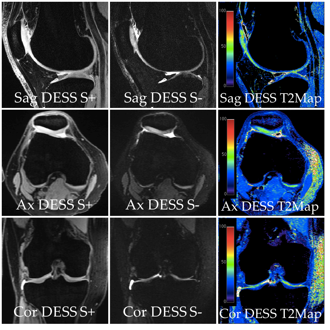
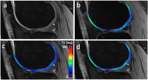
A Simple Analytic Method for Estimating T2 in the Knee from DESS | Body MRI  Research Group (BMR) | Stanford Medicine
A Simple Analytic Method for Estimating T2 in the Knee from DESS | Body MRI Research Group (BMR) | Stanford Medicine
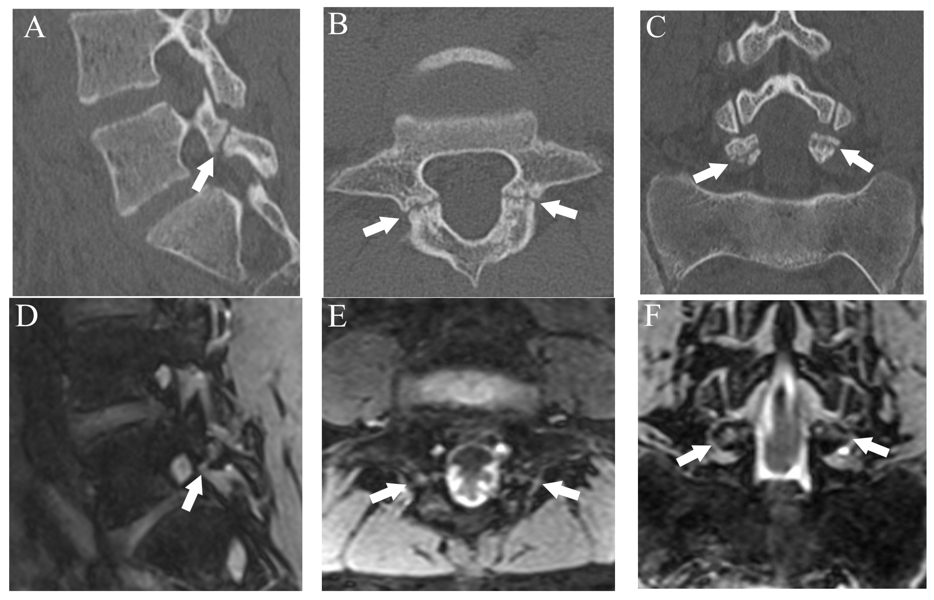
Diagnostics | Free Full-Text | Diagnostic Utility of Double-Echo  Steady-State (DESS) MRI for Fracture and Bone Marrow Edema Detection in  Adolescent Lumbar Spondylolysis
Diagnostics | Free Full-Text | Diagnostic Utility of Double-Echo Steady-State (DESS) MRI for Fracture and Bone Marrow Edema Detection in Adolescent Lumbar Spondylolysis

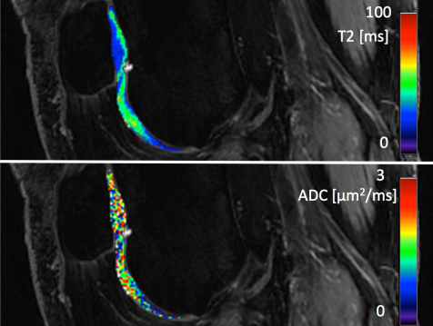
Estimation of Multiple Tissue Parameters Using the DESS Sequence | Body MRI  Research Group (BMR) | Stanford Medicine
Estimation of Multiple Tissue Parameters Using the DESS Sequence | Body MRI Research Group (BMR) | Stanford Medicine

A sample sagittal MRI slice (dualecho steady state (DESS) sequence) of... |  Download Scientific Diagram
A sample sagittal MRI slice (dualecho steady state (DESS) sequence) of... | Download Scientific Diagram

ISMRM2017-000313_Fig2.png

3T MR Imaging of Cartilage using 3D Dual Echo Steady State (DESS)

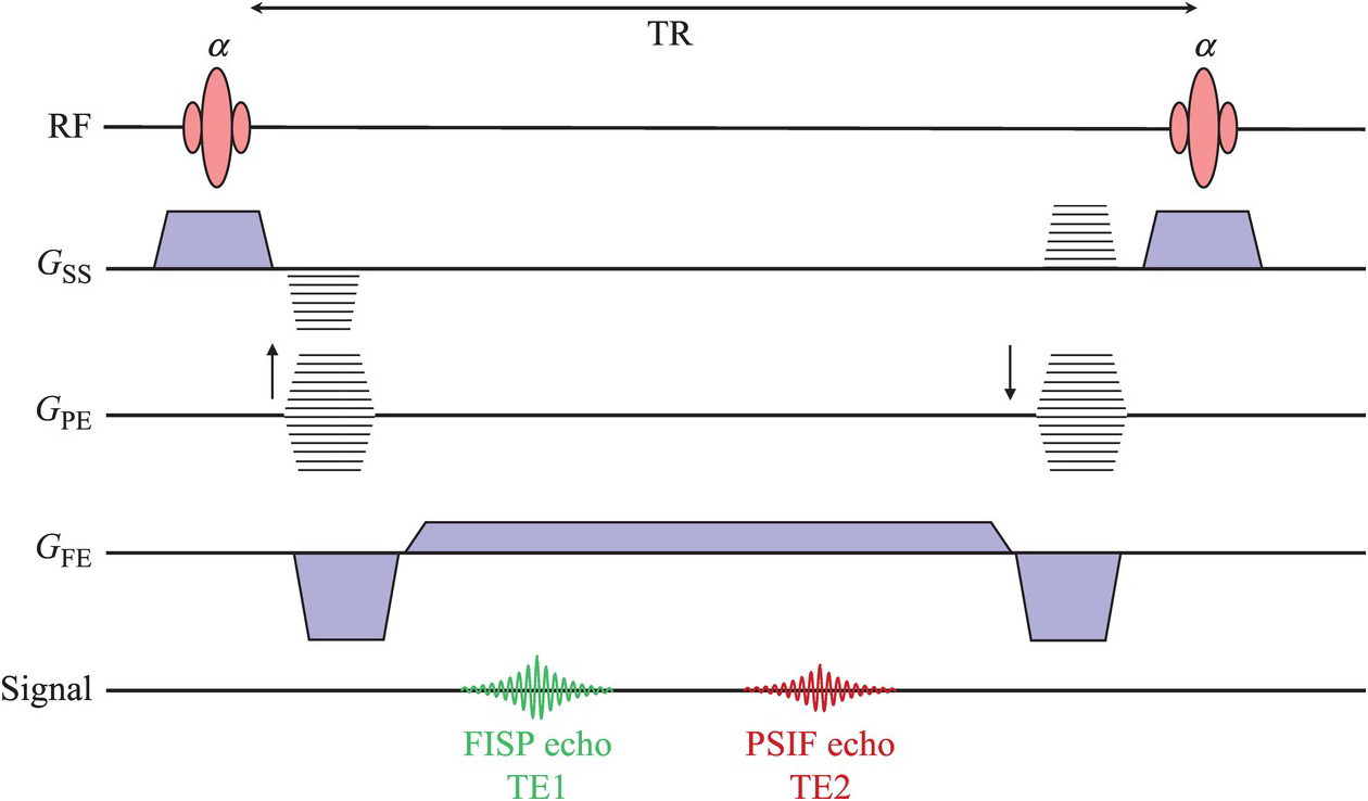
Acronyms Anonymous II: Gradient Echo (Chapter 13) - MRI from Picture to  Proton
Acronyms Anonymous II: Gradient Echo (Chapter 13) - MRI from Picture to Proton

Coronal reconstruction of a DESS sequence showing the lingual nerve... |  Download Scientific Diagram
Coronal reconstruction of a DESS sequence showing the lingual nerve... | Download Scientific Diagram

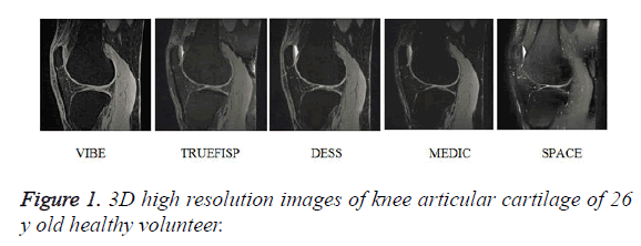
Comparison of 3D MR imaging sequences in knee articular cartilage at 1.5 T

MRI Sequences: T2-enhanced steady-state gradient echo | e-MRI

Double Echo Steady State (DESS) Magnetic Resonance Imaging of Knee  Articular Cartilage at 3 Tesla – a Pilot Study for the Os
Double Echo Steady State (DESS) Magnetic Resonance Imaging of Knee Articular Cartilage at 3 Tesla – a Pilot Study for the Os

PDF] Comparison of 3D MR imaging sequences in knee articular cartilage at  1.5 T | Semantic Scholar
PDF] Comparison of 3D MR imaging sequences in knee articular cartilage at 1.5 T | Semantic Scholar

PDF] Comparison of 3D MR imaging sequences in knee articular cartilage at  1.5 T | Semantic Scholar
PDF] Comparison of 3D MR imaging sequences in knee articular cartilage at 1.5 T | Semantic Scholar

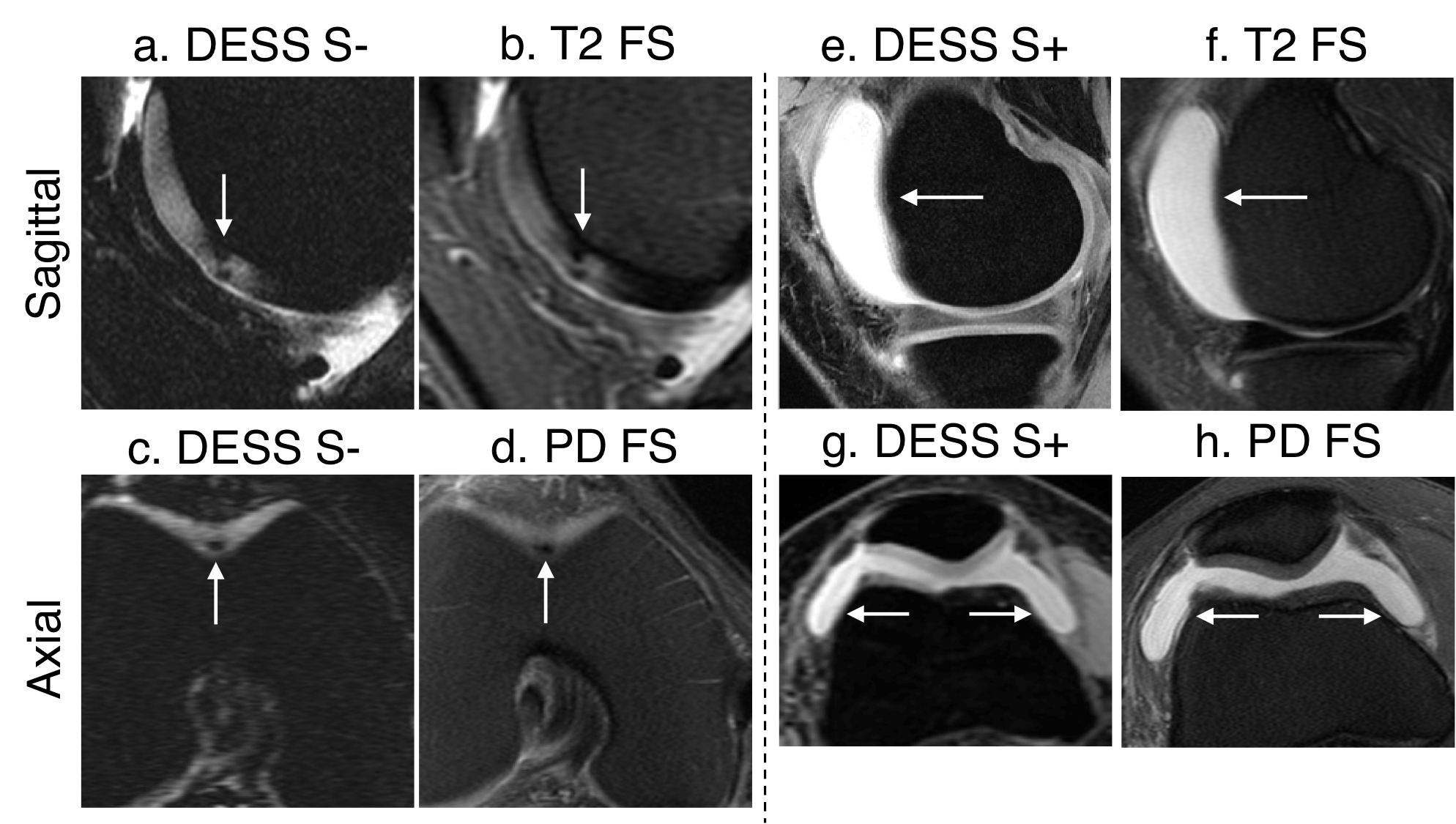
Combined 5‐minute double‐echo in steady‐state with separated echoes and  2‐minute proton‐density‐weighted 2D FSE sequence for comprehensive  whole‐joint knee MRI assessment - Chaudhari - 2019 - Journal of Magnetic  Resonance Imaging - Wiley
Combined 5‐minute double‐echo in steady‐state with separated echoes and 2‐minute proton‐density‐weighted 2D FSE sequence for comprehensive whole‐joint knee MRI assessment - Chaudhari - 2019 - Journal of Magnetic Resonance Imaging - Wiley

3T MR Imaging of Cartilage using 3D Dual Echo Steady State (DESS)

Five‐minute knee MRI for simultaneous morphometry and T2 relaxometry of  cartilage and meniscus and for semiquantitative radiological assessment  using double‐echo in steady‐state at 3T - Chaudhari - 2018 - Journal of  Magnetic
Five‐minute knee MRI for simultaneous morphometry and T2 relaxometry of cartilage and meniscus and for semiquantitative radiological assessment using double‐echo in steady‐state at 3T - Chaudhari - 2018 - Journal of Magnetic

a Coronal and b–d axial reconstruction of the 3D-DESS sequence showing... |  Download Scientific Diagram
a Coronal and b–d axial reconstruction of the 3D-DESS sequence showing... | Download Scientific Diagram

ISMRM2018-000226_Fig5.png

Diagnostics | Free Full-Text | Diagnostic Utility of Double-Echo  Steady-State (DESS) MRI for Fracture and Bone Marrow Edema Detection in  Adolescent Lumbar Spondylolysis
Diagnostics | Free Full-Text | Diagnostic Utility of Double-Echo Steady-State (DESS) MRI for Fracture and Bone Marrow Edema Detection in Adolescent Lumbar Spondylolysis
DESS Sequence (MRI) | Joint and Osteoarthritis Imaging with Novel  Technology Group (JOINT) | Stanford Medicine
DESS Sequence (MRI) | Joint and Osteoarthritis Imaging with Novel Technology Group (JOINT) | Stanford Medicine

Combined 5‐minute double‐echo in steady‐state with separated echoes and  2‐minute proton‐density‐weighted 2D FSE sequence for comprehensive  whole‐joint knee MRI assessment - Chaudhari - 2019 - Journal of Magnetic  Resonance Imaging - Wiley
Combined 5‐minute double‐echo in steady‐state with separated echoes and 2‐minute proton‐density‐weighted 2D FSE sequence for comprehensive whole‐joint knee MRI assessment - Chaudhari - 2019 - Journal of Magnetic Resonance Imaging - Wiley

DESS sequence (25.7/9 ms) with water excitation obtained in a sagittal... |  Download Scientific Diagram
DESS sequence (25.7/9 ms) with water excitation obtained in a sagittal... | Download Scientific Diagram

A and B , Axial source images of the 3D-DESS-WE sequence. C and D ,... |  Download Scientific Diagram
A and B , Axial source images of the 3D-DESS-WE sequence. C and D ,... | Download Scientific Diagram

Rapid T2-mapping in prostate cancer MRI | Dregely Lab