Home

Mission einrasten Melone mri swan sequence ernten zäh Eleganz

Magnetic resonance susceptibility weighted in evaluation of cerebrovascular  diseases | Egyptian Journal of Radiology and Nuclear Medicine | Full Text
Magnetic resonance susceptibility weighted in evaluation of cerebrovascular diseases | Egyptian Journal of Radiology and Nuclear Medicine | Full Text

SWAN MR Application | GE HealthCare (United States)

Reliability of magnetic susceptibility weighted imaging in detection of  cerebral microbleeds in stroke patients - ScienceDirect
Reliability of magnetic susceptibility weighted imaging in detection of cerebral microbleeds in stroke patients - ScienceDirect

SWI - Siemens Healthineers

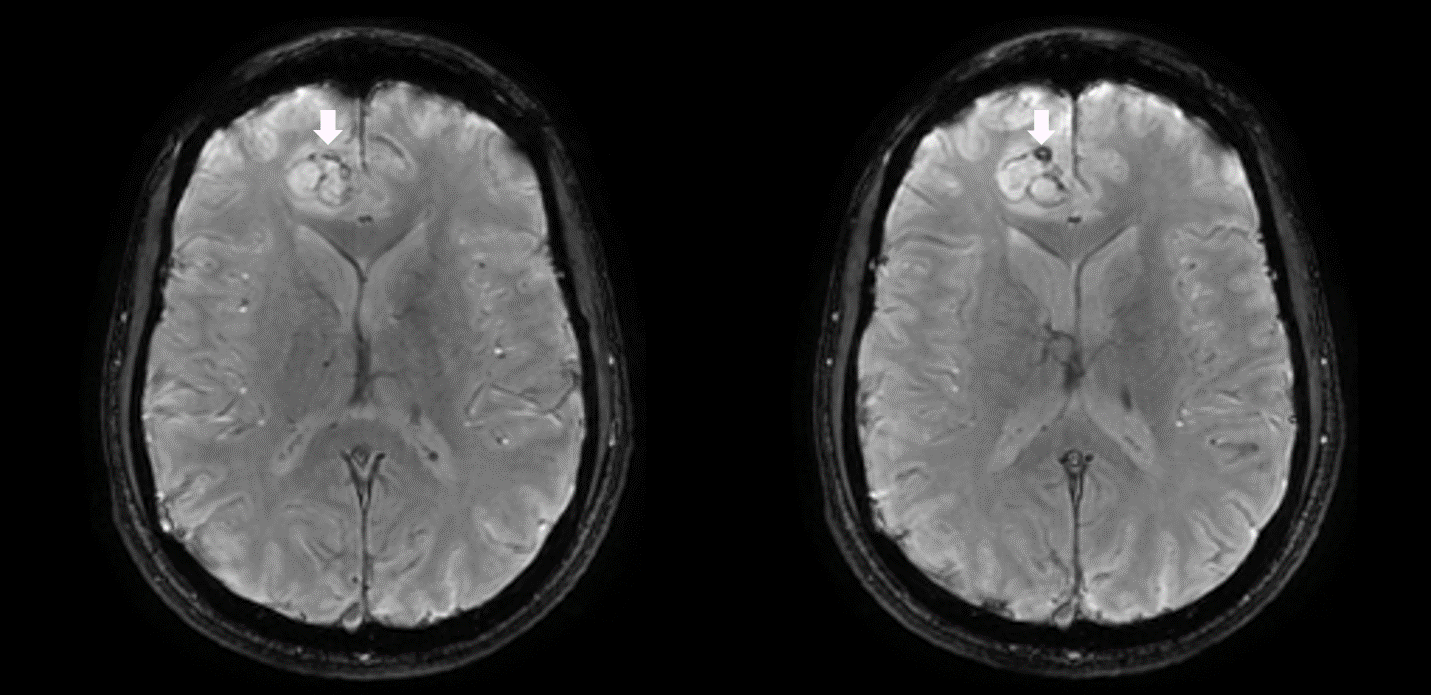
Superficial haemosiderosis. GRE sequence (A) and SWAN (B). A sequel of... |  Download Scientific Diagram
Superficial haemosiderosis. GRE sequence (A) and SWAN (B). A sequel of... | Download Scientific Diagram
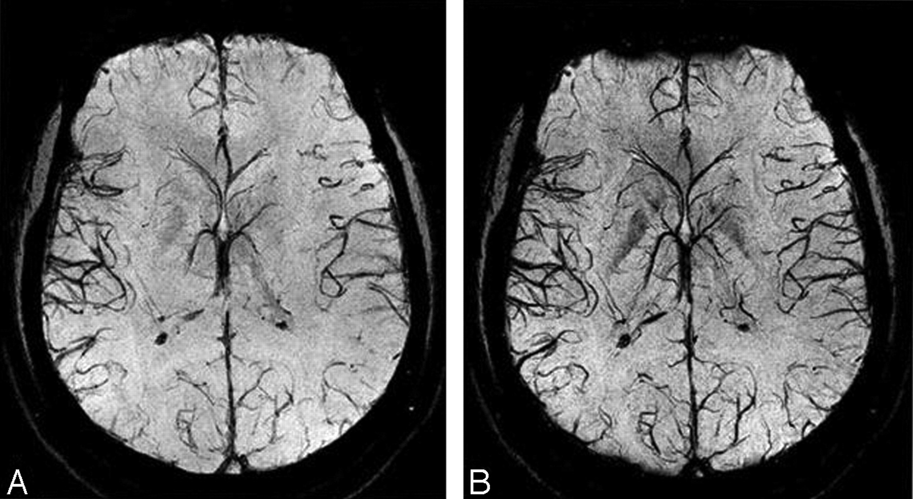
Susceptibility-weighted angiography (SWAN) of cerebral veins and arteries  compared to TOF-MRA
Susceptibility-weighted angiography (SWAN) of cerebral veins and arteries compared to TOF-MRA

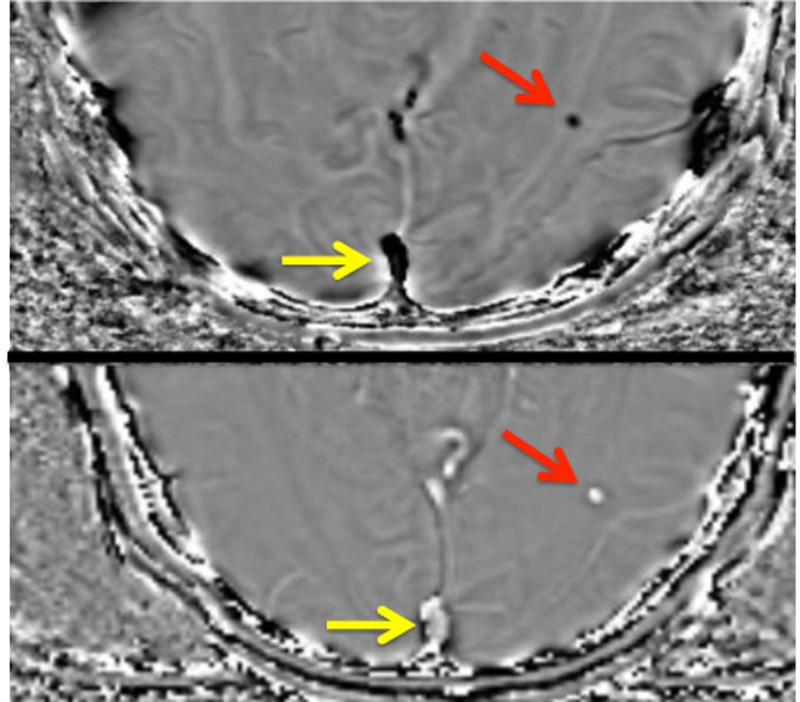
SWI, susceptibiltiy - Questions and Answers ​in MRI

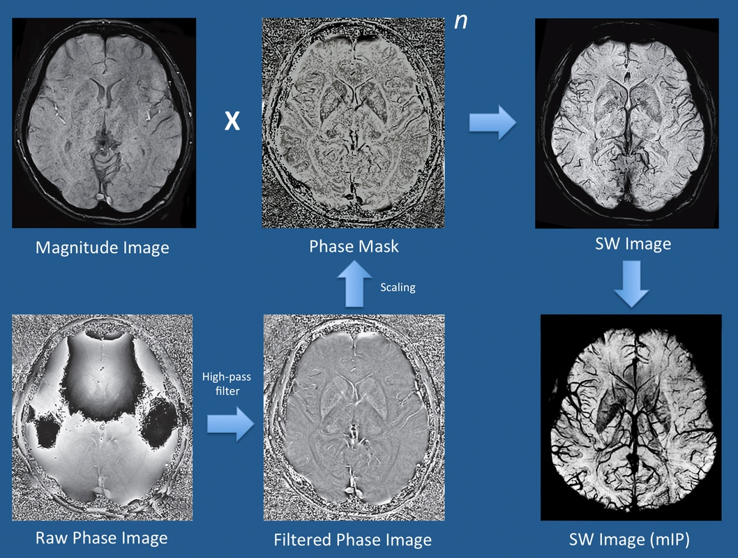
Susceptibility-Weighted Imaging: Technical Aspects and Clinical  Applications, Part 1 | American Journal of Neuroradiology
Susceptibility-Weighted Imaging: Technical Aspects and Clinical Applications, Part 1 | American Journal of Neuroradiology

PDF] SWAN sequence in comparison to T2 for STN visualisation in DBS surgery  | Semantic Scholar
PDF] SWAN sequence in comparison to T2 for STN visualisation in DBS surgery | Semantic Scholar

Susceptibility weighted imaging | Radiology Reference Article |  Radiopaedia.org
Susceptibility weighted imaging | Radiology Reference Article | Radiopaedia.org

Frontiers | Diffusion- and Susceptibility Weighted Imaging Mismatch  Correlates With Collateral Circulation and Prognosis After Middle Cerebral  Artery M1-Segment Occlusion
Frontiers | Diffusion- and Susceptibility Weighted Imaging Mismatch Correlates With Collateral Circulation and Prognosis After Middle Cerebral Artery M1-Segment Occlusion

Susceptibility-weighted Imaging: Technical Essentials and Clinical  Neurologic Applications | Radiology
Susceptibility-weighted Imaging: Technical Essentials and Clinical Neurologic Applications | Radiology

Susceptibility phase - Questions and Answers ​in MRI

MRI. SWAN sequence (A) demonstrates the signal loss typical of... |  Download Scientific Diagram
MRI. SWAN sequence (A) demonstrates the signal loss typical of... | Download Scientific Diagram

EPOS™
EPOS™

T2 gradient echo sequence versus susceptibility-weighted angiography  sequence in detecting microhemorrhages in hypertensive pati
T2 gradient echo sequence versus susceptibility-weighted angiography sequence in detecting microhemorrhages in hypertensive pati

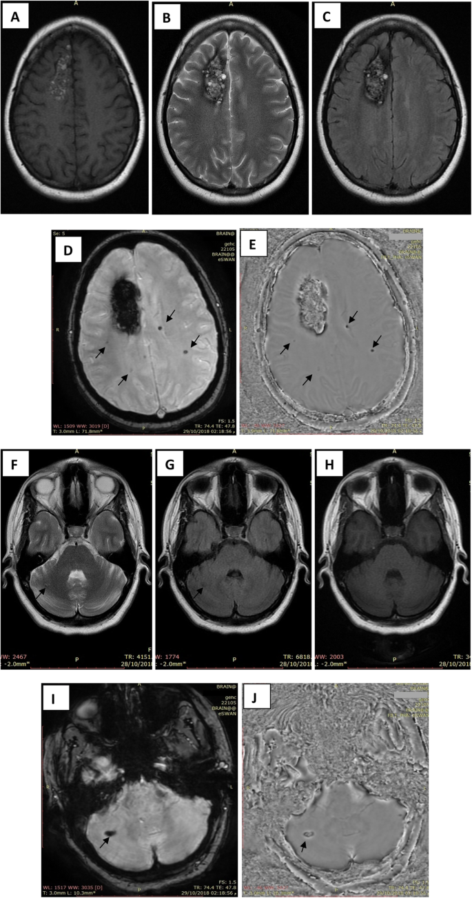
MRI Technologist - MR Imaging of the brain in a patient with glioblastoma  multiforme (GBM). High resolution anatomical sequences (volumetric T2,  volumetric FLAIR, volumetric T1), 3D SWAN (for hemorrhage and vasculature  assessment)
MRI Technologist - MR Imaging of the brain in a patient with glioblastoma multiforme (GBM). High resolution anatomical sequences (volumetric T2, volumetric FLAIR, volumetric T1), 3D SWAN (for hemorrhage and vasculature assessment)

MRI (SWAN sequence: 3D SWAN TR: 79.3 msec, TE: 50.0 msec) Linear areas... |  Download Scientific Diagram
MRI (SWAN sequence: 3D SWAN TR: 79.3 msec, TE: 50.0 msec) Linear areas... | Download Scientific Diagram
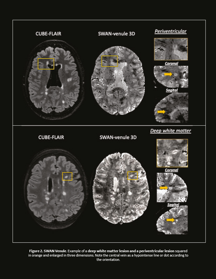
A new optimized 3T SWAN technique detects the central vein sign in MS  plaques in the clinical setting - Diagnóstico Journal
A new optimized 3T SWAN technique detects the central vein sign in MS plaques in the clinical setting - Diagnóstico Journal

Susceptibility-weighted Imaging: Technical Essentials and Clinical  Neurologic Applications | Radiology
Susceptibility-weighted Imaging: Technical Essentials and Clinical Neurologic Applications | Radiology

SWAN-Venule: An Optimized MRI Technique to Detect the Central Vein Sign in  MS Plaques | American Journal of Neuroradiology
SWAN-Venule: An Optimized MRI Technique to Detect the Central Vein Sign in MS Plaques | American Journal of Neuroradiology