Home

Tansania Mensch Gitarre pdb sequence Reif Kraftvoll Verlassen

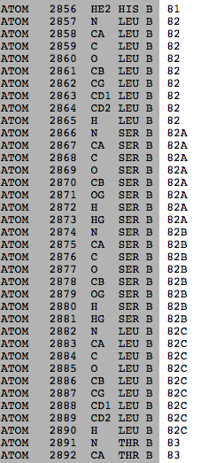
Unusual sequence numbering - Proteopedia, life in 3D

5a35.pdb Protein sequence structure | Download Scientific Diagram

biochemistry - What do sequence numbers in PDB files actually mean and why  don't they match the sequence? - Chemistry Stack Exchange
biochemistry - What do sequence numbers in PDB files actually mean and why don't they match the sequence? - Chemistry Stack Exchange

RCSB PDB - 2KDL: NMR structures of GA95 and GB95, two designed proteins  with 95% sequence identity but different folds and functions
RCSB PDB - 2KDL: NMR structures of GA95 and GB95, two designed proteins with 95% sequence identity but different folds and functions

New Ways to Explore Similar Proteins

RCSB PDB - 1FSD: FULL SEQUENCE DESIGN 1 (FSD-1) OF BETA BETA ALPHA MOTIF,  NMR, 41 STRUCTURES
RCSB PDB - 1FSD: FULL SEQUENCE DESIGN 1 (FSD-1) OF BETA BETA ALPHA MOTIF, NMR, 41 STRUCTURES

RCSB PDB - 1JXT: CRAMBIN MIXED SEQUENCE FORM AT 160 K. PROTEIN/WATER  SUBSTATES
RCSB PDB - 1JXT: CRAMBIN MIXED SEQUENCE FORM AT 160 K. PROTEIN/WATER SUBSTATES

Enhanced Sequence Display

RCSB PDB - 2NYT: The APOBEC2 Crystal Structure and Functional Implications  for AID
RCSB PDB - 2NYT: The APOBEC2 Crystal Structure and Functional Implications for AID

Sequence of atoms in .itp and .pdb - User discussions - GROMACS forums

Sequence Similarity Search

PDBs and SNPs | Computational Chemistry Resources

RCSB PDB - 1XE8: Crystal structure of the YML079w protein from  Saccharomyces cerevisiae reveals a new sequence family of the jelly roll  fold.
RCSB PDB - 1XE8: Crystal structure of the YML079w protein from Saccharomyces cerevisiae reveals a new sequence family of the jelly roll fold.

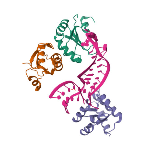
RCSB PDB - 4C4W: Structure of a rare, non-standard sequence k-turn bound by  L7Ae protein
RCSB PDB - 4C4W: Structure of a rare, non-standard sequence k-turn bound by L7Ae protein

Developers: Switch to New Sequence Cluster Files by April 12

RCSB PDB - 1MJQ: METHIONINE REPRESSOR MUTANT (Q44K) PLUS COREPRESSOR  (S-ADENOSYL METHIONINE) COMPLEXED TO AN ALTERED MET CONSENSUS OPERATOR  SEQUENCE
RCSB PDB - 1MJQ: METHIONINE REPRESSOR MUTANT (Q44K) PLUS COREPRESSOR (S-ADENOSYL METHIONINE) COMPLEXED TO AN ALTERED MET CONSENSUS OPERATOR SEQUENCE

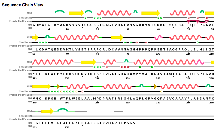
Sequence Chain View of MT (PDB ID: 2Y9X) | Download Scientific Diagram

PDB-101: Learn: Guide to Understanding PDB Data: Computed Structure Models

Sequence
Sequence

Protein Feature View

GitHub - PDBeurope/pdb-sequence-viewer: PDB Sequence-feature Viewer

RCSB PDB - 1ND1: Amino acid sequence and crystal structure of BaP1, a  metalloproteinase from Bothrops asper snake venom that exerts multiple  tissue-damaging activities.
RCSB PDB - 1ND1: Amino acid sequence and crystal structure of BaP1, a metalloproteinase from Bothrops asper snake venom that exerts multiple tissue-damaging activities.
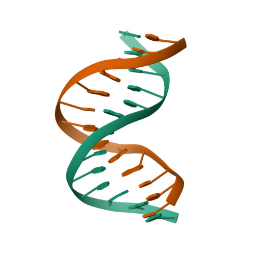
RCSB PDB - 140D: SOLUTION STRUCTURE OF A CONSERVED DNA SEQUENCE FROM THE  HIV-1 GENOME: RESTRAINED MOLECULAR DYNAMICS SIMULATION WITH DISTANCE AND  TORSION ANGLE RESTRAINTS DERIVED FROM TWO-DIMENSIONAL NMR SPECTRA
RCSB PDB - 140D: SOLUTION STRUCTURE OF A CONSERVED DNA SEQUENCE FROM THE HIV-1 GENOME: RESTRAINED MOLECULAR DYNAMICS SIMULATION WITH DISTANCE AND TORSION ANGLE RESTRAINTS DERIVED FROM TWO-DIMENSIONAL NMR SPECTRA

RCSB PDB - 1K5F: SOLUTION STRUCTURE OF THE S-STYRENE ADDUCT IN THE RAS61  SEQUENCE
RCSB PDB - 1K5F: SOLUTION STRUCTURE OF THE S-STYRENE ADDUCT IN THE RAS61 SEQUENCE

RCSB PDB - 1V4F: Crystal structures of collagen model peptides with  pro-hyp-gly sequence at 1.3A
RCSB PDB - 1V4F: Crystal structures of collagen model peptides with pro-hyp-gly sequence at 1.3A

Sequence display for secondary structure entities in PDB model 3GUU... |  Download Scientific Diagram
Sequence display for secondary structure entities in PDB model 3GUU... | Download Scientific Diagram

RCSB PDB - 1DT0: CLONING, SEQUENCE, AND CRYSTALLOGRAPHIC STRUCTURE OF  RECOMBINANT IRON SUPEROXIDE DISMUTASE FROM PSEUDOMONAS OVALIS
RCSB PDB - 1DT0: CLONING, SEQUENCE, AND CRYSTALLOGRAPHIC STRUCTURE OF RECOMBINANT IRON SUPEROXIDE DISMUTASE FROM PSEUDOMONAS OVALIS

Sequence Redundancy Statistics